الأسبوع 1 : Jaundice
2 مشترك
طلاب 2005 بكلية الطب الجامعة الاردنية :: أرشيف سنة رابعة :: جراحة :: الجراحة surgery :: موضوع الأسبوع
صفحة 1 من اصل 1

الأسبوع 1 : Jaundice
من طرف Admin الأحد أكتوبر 19, 2008 11:28 am
Jaundice
السلام عليكم
اول أسبوع سوف يكون موضوع النقاش عن ال Jaundice
فأتمنى من الجميع المشاركة والنقاش
ووضع مقالات من الانترنت و صور وفيديو او حتى معلومات بلغة العضو نفسه
بالاضافة لطرح استفسارات والاجابة عليها
سيتم مناقشة هذا الموضوع حتى الأحد القادم باذن الله
السلام عليكم
اول أسبوع سوف يكون موضوع النقاش عن ال Jaundice
فأتمنى من الجميع المشاركة والنقاش
ووضع مقالات من الانترنت و صور وفيديو او حتى معلومات بلغة العضو نفسه
بالاضافة لطرح استفسارات والاجابة عليها
سيتم مناقشة هذا الموضوع حتى الأحد القادم باذن الله

Admin- Admin
- عدد المساهمات : 443
تاريخ التسجيل : 11/10/2008 -

رد: الأسبوع 1 : Jaundice
من طرف Admin الأحد أكتوبر 19, 2008 4:41 pm
Jaundice
, also known as icterus (attributive adjective: "icteric"), is yellowish discoloration of the skin, sclerae (whites of the eyes) and mucous membranes caused by hyperbilirubinemia (increased levels of bilirubin in the blood). This hyperbilirubinemia subsequently causes increased levels of bilirubin in the extracellular fluids. Typically, the concentration of bilirubin in the plasma must exceed 1.5 mg/dL[1], three times the usual value of approximately 0.5mg/dL[1], for the coloration to be easily visible. Jaundice comes from the French word jaune, meaning yellow.
* 1 Normal Physiology
o 1.1 Pre-hepatic events
o 1.2 Hepatic events
o 1.3 Post-hepatic events
* 2 Causes
o 2.1 Pre-hepatic
o 2.2 Hepatic
o 2.3 Post-hepatic
o 2.4 Laboratory Tests
* 3 Neonatal jaundice
* 4 Jaundiced eye
Normal Physiology
In order to understand how jaundice results, it is important to understand where the pathological processes that cause jaundice take their effect. It is also important to further recognize that jaundice itself is not a disease, but rather a symptom of an underlying pathological process that occurs at some point along the normal physiological pathway of the metabolism of bilirubin.
Pre-hepatic events
When red blood cells have completed their life span of approximately 120 days, or when they are damaged, their membranes become fragile and prone to rupture. As the cell traverses through the reticuloendothelial system, their cell membranes rupture and the contents of the red blood cell is released into the blood. The component of the red blood cell that is involved in jaundice is hemoglobin. The hemoglobin released into the blood is phagocytosed by macrophages, and split into its heme and globin portions. The globin portion, being protein, is degraded into amino acids and plays no further role in jaundice. Two reactions then take place to the heme molecule. The first reaction is the oxidation of heme to form biliverdin.This reaction is catalyzed by microsomal enzyme heme oxygenase and it results in biliverdin (green color pigment), iron and carbon monoxide. Next step is reduction of biliverdin to yellow color tetrapyrol pigment bilirubin by cytosolic enzyme biliverdin reductase. This bilirubin is known as "unconjugated", "free" or "indirect" bilirubin. Approximately 4 mg per kg of bilirubin is produced each day.[2] The majority of this bilirubin comes from the breakdown of heme from expired red blood cells in the process just described. However approximately 20 per cent comes from other heme sources, including ineffective erythropoiesis, breakdown of other heme-containing proteins, such as muscle myoglobin and cytochromes.[2]
Hepatic events
The unconjugated bilirubin then travels to the liver through the bloodstream. Because this bilirubin is not soluble, however, it is transported through the blood bound to serum albumin. Once it arrives at the liver, it is conjugated with glucuronic acid (to form bilirubin diglucuronide, or just "conjugated bilirubin") to become more water soluble. The reaction is catalyzed by the enzyme UDP-glucuronide transferase.
Post-hepatic events
This conjugated bilirubin is excreted from the liver into the biliary and cystic ducts as part of bile. Intestinal bacteria convert the bilirubin into urobilinogen. From here the urobilinogen can take two pathways. It can either be further converted into stercobilinogen, which is then oxidized to stercobilin and passed out in the faeces, or it can be reabsorbed by the intestinal cells, transported in the blood to the kidneys, and passed out in the urine as the oxidised product urobilin. Stercobilin and urobilin are the products responsible for the coloration of faeces and urine, respectively.
Causes
When a pathological process interferes with the normal functioning of the metabolism and excretion of bilirubin just described, jaundice may be the result. Jaundice is classified into three categories, depending on which part of the physiological mechanism the pathology affects. The three categories are:
* Pre-hepatic: The pathology is occurring prior the liver
* Hepatic: The pathology is located within the liver
* Post-Hepatic: The pathology is located after the conjugation of bilirubin in the liver
Pre-hepatic
Pre-hepatic jaundice is caused by anything which causes an increased rate of hemolysis (breakdown of red blood cells). In tropical countries, malaria can cause jaundice in this manner. Certain genetic diseases, such as sickle cell anemia, spherocytosis and glucose 6-phosphate dehydrogenase deficiency can lead to increased red cell lysis and therefore hemolytic jaundice. Commonly, diseases of the kidney, such as hemolytic uremic syndrome, can also lead to coloration. Defects in bilirubin metabolism also present as jaundice. Jaundice usually comes with high fevers.
Laboratory findings include:
* Urine: no bilirubin present, urobilirubin > 2 units (except in infants where gut flora has not developed).
* Serum: increased unconjugated bilirubin.
Hepatic
Hepatic jaundice causes include acute hepatitis, hepatotoxicity and alcoholic liver disease, whereby cell necrosis reduces the liver's ability to metabolise and excrete bilirubin leading to a buildup in the blood. Less common causes include primary biliary cirrhosis, Gilbert's syndrome (a genetic disorder of bilirubin metabolism which can result in mild jaundice, which is found in about 5% of the population), Crigler-Najjar syndrome, metastatic carcinoma and Niemann Pick Type C disease. Jaundice seen in the newborn, known as neonatal jaundice, is common, occurring in almost every newborn as hepatic machinery for the conjugation and excretion of bilirubin does not fully mature until approximately two weeks of age.
Laboratory Findings include:
* Urine: Conjugated bilirubin present, Urobilirubin > 2 units but variable (Except in children)
Post-hepatic
Post-hepatic jaundice, also called obstructive jaundice, is caused by an interruption to the drainage of bile in the biliary system. The most common causes are gallstones in the common bile duct, and pancreatic cancer in the head of the pancreas. Also, a group of parasites known as "liver flukes" live in the common bile duct, causing obstructive jaundice. Other causes include strictures of the common bile duct, biliary atresia, ductal carcinoma, pancreatitis and pancreatic pseudocysts. A rare cause of obstructive jaundice is Mirizzi's syndrome.
The presence of pale stools and dark urine suggests an obstructive or post-hepatic cause as normal feces get their color from bile pigments.
Patients also can present with elevated serum cholesterol, and often complain of severe itching or "pruritus".
Laboratory Tests
No one test can differentiate between various classifications of jaundice. A combinations of liver function tests is essential to
arrive at a diagnosis.
Neonatal jaundice
Neonatal jaundice is usually harmless: this condition is often seen in infants around the second day after birth, lasting until day 8 in normal births, or to around day 14 in premature births. Serum bilirubin normally drops to a low level without any intervention required: the jaundice is presumably a consequence of metabolic and physiological adjustments after birth. In extreme cases, a brain-damaging condition known as kernicterus can occur; there are concerns that this condition has been rising in recent years due to inadequate detection and treatment of neonatal hyperbilirubinemia. Neonatal jaundice is a risk factor for hearing loss.[3]
Jaundiced eye
It was once believed persons suffering from the medical condition jaundice saw everything as yellow. By extension, the jaundiced eye came to mean a prejudiced view, usually rather negative or critical. Alexander Pope, in 'An Essay on Criticism' (1711), wrote: "All seems infected that the infected spy, As all looks yellow to the jaundiced eye."[4] Similarly in the mid 19th century the English poet Lord Alfred Tennyson wrote in the poem 'Locksley Hall': "So I triumphe'd ere my passion sweeping thro' me left me dry, left me with the palsied heart, and left me with a jaundiced eye."
عدل سابقا من قبل Admin في الأحد أكتوبر 19, 2008 4:47 pm عدل 1 مرات
Admin- Admin
- عدد المساهمات : 443
تاريخ التسجيل : 11/10/2008 -
رد: الأسبوع 1 : Jaundice
من طرف Admin الأحد أكتوبر 19, 2008 4:43 pm
Yellowing of the skin and sclera caused by Hepatitis A.
Admin- Admin
- عدد المساهمات : 443
تاريخ التسجيل : 11/10/2008 -
رد: الأسبوع 1 : Jaundice
من طرف اسراء الأحد أكتوبر 19, 2008 7:54 pm

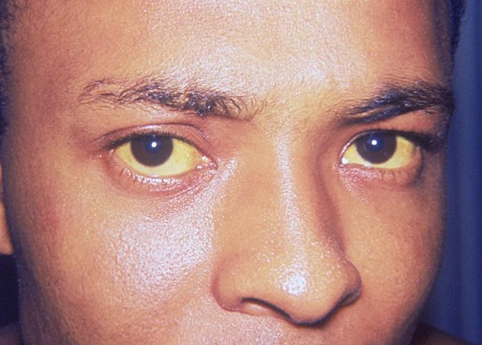
Jaundice in a male with hepatic failure


اسراء- عدد المساهمات : 694
تاريخ التسجيل : 19/10/2008
العمر : 36 -
رد: الأسبوع 1 : Jaundice
من طرف اسراء الأحد أكتوبر 19, 2008 8:09 pm
اسراء- عدد المساهمات : 694
تاريخ التسجيل : 19/10/2008
العمر : 36 -
رد: الأسبوع 1 : Jaundice
من طرف اسراء الأحد أكتوبر 19, 2008 8:16 pm
How is the cause of jaundice diagnosed?
Many tests are available for determining the cause of jaundice, but the history and physical examination are important as well.
History
The history can suggest possible reasons for the jaundice. For example, heavy use of alcohol suggests alcoholic liver disease, whereas use of illegal, injectable drugs suggests viral hepatitis. Recent initiation of a new drug suggests drug-induced jaundice. Episodes of abdominal pain associated with jaundice suggests blockage of the bile ducts usually by gallstones.
Physical examination
The most important part of the physical examination in a patient who is jaundiced is examination of the abdomen. Masses (tumors) in the abdomen suggest cancer infiltrating the liver (metastatic cancer) as the cause of the jaundice. An enlarged, firm liver suggests cirrhosis. A rock-hard, nodular liver suggests cancer within the liver.
Blood tests
Measurement of bilirubin can be helpful in determining the causes of jaundice. Markedly greater elevations of unconjugated bilirubin relative to elevations of conjugated bilirubin in the blood suggest hemolysis (destruction of red blood cells). Marked elevations of liver tests (aspartate amino transferase or AST and alanine amino transferase or ALT) suggest inflammation of the liver (such as viral hepatitis). Elevations of other liver tests, e.g., alkaline phosphatase, suggest diseases or obstruction of the bile ducts.
Ultrasonography
Ultrasonography is a simple, safe, and readily-available test that uses sound waves to examine the organs within the abdomen. Ultrasound examination of the abdomen may disclose gallstones, tumors in the liver or the pancreas, and dilated bile ducts due to obstruction (by gallstones or tumor).
Computerized tomography (CT or CAT scans)
Computerized tomography or CT scans are scans that use x-rays to examine the soft tissues of the abdomen. They are particularly good for identifying tumors in the liver and the pancreas and dilated bile ducts, though they are not as good as ultrasonography for identifying gallstones.
Magnetic resonance imaging (MRI)
Magnetic Resonance Imaging scans are scans that utilize magnetization of the body to examine the soft tissues of the abdomen. Like CT scans, they are good for identifying tumors and studying bile ducts. MRI scans can be modified to visualize the bile ducts better than CT scans (a procedure referred to as MR cholangiography), and, therefore, are better than CT for identifying the cause and location of bile duct obstruction.
Endoscopic retrograde cholangiopancreatography (ERCP) and endoscopic ultrasound
Endoscopic retrograde cholangiopancreatography (ERCP) provides the best means for examining the bile duct. For ERCP an endoscope is swallowed by the patient after he or she has been sedated. The endoscope is a flexible, fiberoptic tube approximately four feet in length with a light and camera on its tip. The tip of the endoscope is passed down the esophagus, through the stomach, and into the duodenum where the main bile duct enters the intestine. A thin tube then is passed through the endoscope and into the bile duct, and the duct is filled with x-ray contrast solution. An x-ray is taken that clearly demonstrates the contrast-filled bile ducts. ERCP is particularly good at demonstrating the cause and location of obstruction within the bile ducts. A major advantage of ERCP is that diagnostic and therapeutic procedures can be done at the same time as the x-rays. For example, if gallstones are found in the bile ducts, they can be removed. Stents can be placed in the bile ducts to relieve the obstruction caused by scarring or tumors. Biopsies of tumors can be obtained.
Ultrasonography can be combined with ERCP by using a specialized endoscope capable of doing ultrasound scanning. Endoscopic ultrasound is excellent for diagnosing small gallstones in the gallbladder and bile ducts that can be missed by other diagnostic methods such as ultrasound, CT, and MRI. It also is the best means of examining the pancreas for tumors and can facilitate biopsy through the endoscope of tumors within the pancreas.
Liver biopsy
Biopsy of the liver provides a small piece of tissue from the liver for examination under the microscope. The biopsy most commonly is done with a long needle after local injection of the skin of the abdomen overlying the liver with anesthetic. The needle passes through the skin and into the liver, cutting off a small piece of liver tissue. When the needle is withdrawn, the piece of liver comes with it. Liver biopsy is particularly good for diagnosing inflammation of the liver and bile ducts, cirrhosis, cancer, and fatty liver.
How is jaundice treated?
With the exception of the treatments for specific causes of jaundice mentioned previously, the treatment of jaundice usually requires a diagnosis of the specific cause of the jaundice and treatment directed at the specific cause, e.g., removal of a gallstone blocking the bile duct.
اسراء- عدد المساهمات : 694
تاريخ التسجيل : 19/10/2008
العمر : 36 -

Admin- Admin
- عدد المساهمات : 443
تاريخ التسجيل : 11/10/2008 -
طلاب 2005 بكلية الطب الجامعة الاردنية :: أرشيف سنة رابعة :: جراحة :: الجراحة surgery :: موضوع الأسبوع
صفحة 1 من اصل 1
صلاحيات هذا المنتدى:
لاتستطيع الرد على المواضيع في هذا المنتدى