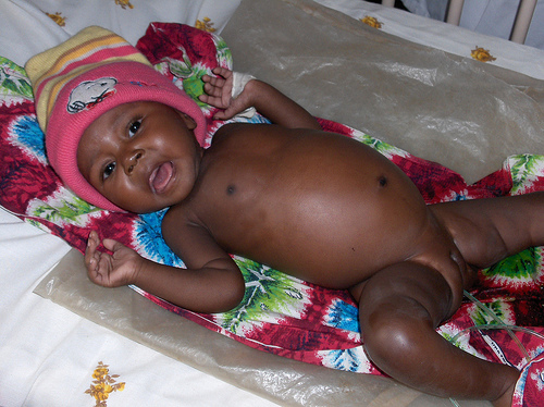
Pediatric Images & Vedios
3 مشترك
طلاب 2005 بكلية الطب الجامعة الاردنية :: أرشيف سنة رابعة :: جراحة :: الجراحة surgery :: الوسائط : الصور و الفيديو
صفحة 1 من اصل 1
 Pediatric Images & Vedios
السلام عليكم
سوف نخصص هذا الموضوع لصور وفيديوهات خاصة في مادة الاطفال مع نبذه صغيرة عن الموضوع وال findings
سوف نخصص هذا الموضوع لصور وفيديوهات خاصة في مادة الاطفال مع نبذه صغيرة عن الموضوع وال findings
INTESTINAL OBSTRUCTION:
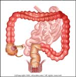
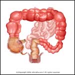
* Large bowel (colon) obstruction may occur as a result of the following conditions:
1. Intrinsic cancer of the colon (70% of all cases)
2. Inflammatory strictures
3. Extrinsic lesions such as a hernia or a neoplasm (rapped outside the intestine)
4. Intussusception
5. Hirschsprung's disease
6. Post abdominal surgery adhesions
7. Fecal Impaction
8. Secondary to volvulus and Diverticulitis
* Obstruction of the left side of the colon is more common.
* Treatment is geared to the underlying cause.


Figure 1 shows a normal abdomen. Figure 2 shows air trapped in the bowels because gas, fluids, or solids cannot move through the bowels normally.
?????- زائر
 رد: Pediatric Images & Vedios
مشكورة اخت اسراء والصور و المعلومات مفيدة جدا
اتمنى تستمري في هذا الموضوع ومواضيع اخرى
اتمنى تستمري في هذا الموضوع ومواضيع اخرى
 رد: Pediatric Images & Vedios
Admin كتب:مشكورة اخت اسراء والصور و المعلومات مفيدة جدا
اتمنى تستمري في هذا الموضوع ومواضيع اخرى
العفو
ان شاء الله نقدر نساعد معكم
والله يقويكم
 رد: Pediatric Images & Vedios
ما شاء الله
رائع دكتورة
رائع دكتورة

Scorpion Payne- عدد المساهمات : 266
تاريخ التسجيل : 18/10/2008
العمر : 37
 رد: Pediatric Images & Vedios
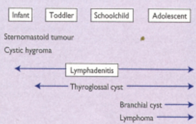
LUMPS AND SWELLINGS OF THE HEAD AND NECK IN CHILDREN
Commonest causes of neck lumps in children by age.
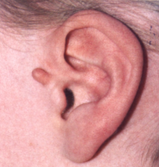
Preauricular cartilaginous remnant
Bilateral branchial sinuses, related to anterior border of sternomastoid.

Thyroglossal cyst--exaggerated by neck extension.
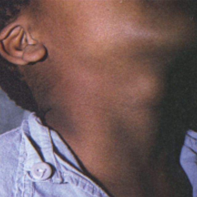
Cystic hygroma arising from posterior triangle of neck.

Commonest causes of neck lumps in children by age.

Preauricular cartilaginous remnant

Bilateral branchial sinuses, related to anterior border of sternomastoid.

Thyroglossal cyst--exaggerated by neck extension.

Cystic hygroma arising from posterior triangle of neck.

 رد: Pediatric Images & Vedios

Clinical findings of case 4.
(A) Voiding cystourethrography (VCG), showing left vesicoureteral reflux (VUR; grade 3) and hydronephrosis.
لاحظ توسع الحالب الى الاعلى على الجهة اليسرى مقارنة باليمنى
(B) External genitalia, with micropenis, bifid scrotum, hypospadias, and undescended testes.
 رد: Pediatric Images & Vedios
ECTOPIC KIDNEY

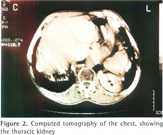


This female patient underwent routine abdominal ultrasound imaging. These ultrasound images show the right kidney just to the right of the uterus in the true pelvis. There was no kidney visualized in the right renal fossa. These sonographic images are diagnostic of ectopic kidney (in this case, a pelvic kidney). It is unusual to see the pelvic kidney so low down in the pelvis


This female patient underwent routine abdominal ultrasound imaging. These ultrasound images show the right kidney just to the right of the uterus in the true pelvis. There was no kidney visualized in the right renal fossa. These sonographic images are diagnostic of ectopic kidney (in this case, a pelvic kidney). It is unusual to see the pelvic kidney so low down in the pelvis
 مواضيع مماثلة
» Acute Gastritis vedios
» سلايدات الدكتور محمد العمري-PEDIATRIC
» سلايدات الدكتور حسان الجديد-Pediatric
» سلايدات الدكتور محمد العمري-PEDIATRIC
» سلايدات الدكتور حسان الجديد-Pediatric
طلاب 2005 بكلية الطب الجامعة الاردنية :: أرشيف سنة رابعة :: جراحة :: الجراحة surgery :: الوسائط : الصور و الفيديو
صفحة 1 من اصل 1
صلاحيات هذا المنتدى:
لاتستطيع الرد على المواضيع في هذا المنتدى
